MACS Matchmaker
The NHS-adapter strategy enables covalent, high-density immobilization of unmodified, primary-amine-bearing ligands directly on an Oligo|PEG mologram. Unlike off-chip DDI, no oligo conjugation kit is required — the NHS-equivalent reactive group is presented through a bifunctional linker that is loaded onto the chip in situ. The duplex underneath stays the weakest link, so the surface is regenerable between ligands by alkaline dehybridization.
Before you start
- Read Safety & Compliance and the safety data sheet for every reagent below. The SDS, not this page, governs how a reagent is handled, stored and disposed of.
- Use the personal protective equipment and the waste route your laboratory prescribes for these reagent classes.
- Check this protocol against the materials actually in hand: confirm the intended Seq ID, the product revision, the lot-specific certificate and the instructions supplied with it. Where those differ from this page, the documentation supplied with the material governs.
From the catalogue
Order the Oligo Adapter NHS directly from the lino Biotech webshop:
- Oligo Adapter NHS — 110 µL stock at 2.6 µM in PBST, 10 immobilizations at 200 nM (130 µL/injection), pre-conjugated to your chosen Seq ID (01–64) for multiplexing on Oligo|PEG (8×8 default; 6×9 on request). Keep the TCO–PEG–TFP linker on hand separately and reconstitute it fresh in MES pH 6.0 before each use.
Strategy: a three-component adapter system
Coupling proceeds in three loading steps on an Oligo|PEG sensor:
- Hybridise the Me-Tz-oligo adapter onto the chip, presenting tetrazine groups on every active ridge.
- Click the bifunctional linker (TCO–PEG–TFP) at mildly acidic pH. The TCO end reacts with the surface tetrazine via inverse-electron-demand Diels–Alder (IEDDA); the TFP / NHS ester points outwards, ready to couple primary amines.
- Inject the ligand. Surface lysines of the ligand react with the exposed TFP / NHS ester to form a stable amide bond, locking the ligand covalently onto the activated ridges.
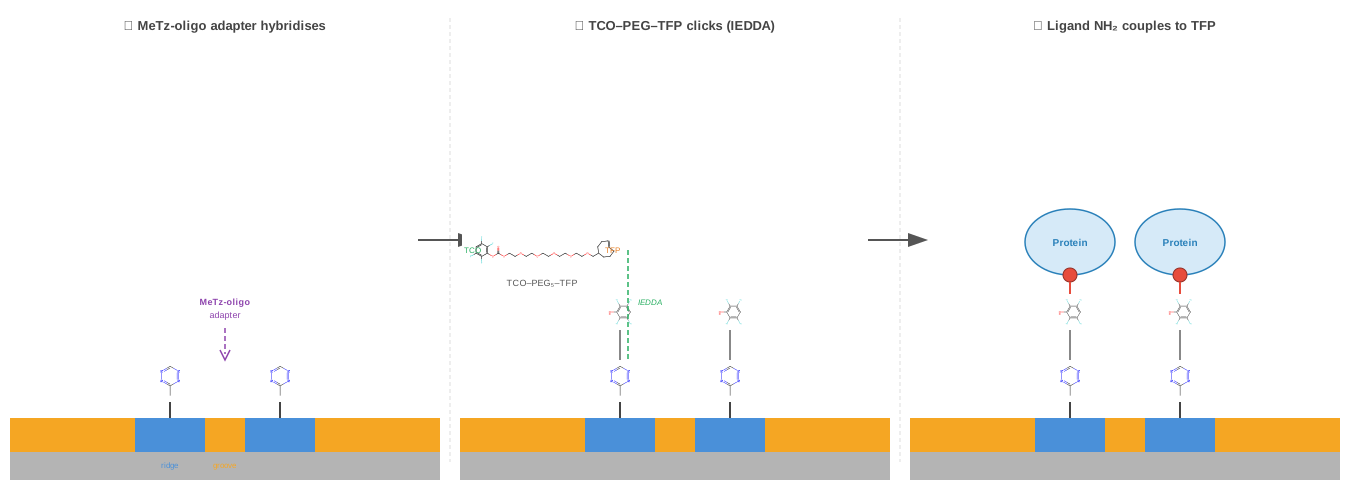
A short ethanolamine wash quenches any residual TFP / NHS groups before analyte injection, and an optional alkaline regeneration step removes non-covalently bound analyte for ligand reuse.
Protocol
Step 1 — Sensor
Use any Oligo|PEG sensor (1- to 64-plex). Multiplexed formats are supported.
Step 2 — Adapter hybridization
| Parameter | Value |
|---|---|
| Reagent | Me-Tz-functionalized oligo adapter, complementary to the chip oligo |
| Concentration | 200 nM |
| Buffer | PBST (phosphate-buffered saline + 0.05% Tween 20) |
| Flow rate | 10 µL/min |
Step 3 — Linker activation (TCO–tetrazine click)
| Parameter | Value |
|---|---|
| Reagent | TCO–PEG5–TFP (bifunctional linker) |
| Linker chemistry preference | TFP > STP > NHS (slower hydrolysis = higher coupling yield; see Hermanson 2013) |
| Concentration | 20–50 µM (optimal); up to 100 µM |
| Buffer | MES pH 6.0 + 0.05% Tween 20 |
| Flow rate | 10 µL/min |
| Incubation | 5 min |
Step 4 — Ligand immobilization
| Parameter | Value |
|---|---|
| Ligand | Any primary-amine-bearing protein or peptide (IgG, enzymes, etc.) |
| Concentration | 0.1–0.5 µM |
| Buffer | HEPES pH 7.0 + 0.05% Tween 20 |
| Flow rate | 10 µL/min |
| Expected coverage | ~190 pg/mm² covalent immobilization under benchmark conditions |
Step 5 — Passivation
| Parameter | Value |
|---|---|
| Reagent | Ethanolamine, 1 M, pH 8.5 |
| Flow rate | 10 µL/min |
| Incubation | 5 min |
| Purpose | Quenches unreacted TFP / NHS esters on the activated molograms |
Step 6 — Analyte injection
| Parameter | Value |
|---|---|
| SCK association flow rate | 30 µL/min (6-ch) / 60 µL/min (3–4-ch) / 100 µL/min (1-ch) |
| SCK dissociation flow rate | 200 µL/min |
Step 7 — Optional regeneration
An alkaline wash removes non-covalently bound analyte while leaving the covalently attached ligand in place — the chip can be re-used with fresh analyte injections. For full ligand replacement, dehybridize the duplex underneath and start again from Step 2.
Reference protocol parameters
| Parameter | Reference value |
|---|---|
| Adapter | Me-Tz-functionalized oligo (preferred over TCO-oligo) |
| Linker | TCO–PEG5–TFP |
| Linker concentration | 20–50 µM |
| Linker buffer | MES pH 6.0 + 0.05% Tween 20, 5 min |
| Ligand concentration | 0.1–0.5 µM |
| Ligand buffer | HEPES pH 7.0 + 0.05% Tween 20 |
| Passivation | 1 M ethanolamine pH 8.5, 5 min |
| On-chip flow rate (all loading steps) | 10 µL/min |
| SCK association flow rate | 30 µL/min (6-ch) / 60 µL/min (3–4-ch) / 100 µL/min (1-ch) |
| SCK dissociation flow rate | 200 µL/min |
| Tween 20 | 0.05% in linker, passivation, and regeneration buffers |
| Ionic strength | Low (PBST or MES-buffered saline) to enable surface pre-concentration |
QC and acceptance
Run these three checks after passivation and before the first analyte injection. Reaching an analyte injection on a surface that has not passed them wastes the sample and produces a result that cannot be interpreted either way.
- Ligand is on the surface. The ligand injection rose and the response persisted through the following wash. A response that decays back toward baseline was pre-concentration without coupling.
- The baseline returns. A buffer injection comes back to the pre-injection level. Upward drift here is adsorption to the PEG grooves and will be read as binding later.
- The surface is selective. A non-target protein at a comparable concentration produces no coherent response.
Passing all three establishes that the chemistry worked; it does not establish that the ligand is active. That needs the analyte, or a known-active control ligand alongside it.
Practical notes
Looking for the broader chip + adapter selection? Start from the interactive decision tree — the NHS on-chip lysine-coupling branch lands here.
Troubleshooting
Symptom: low ligand coverage, weak ligand signal
The most common failure mode of the NHS-adapter protocol is poor electrostatic pre-concentration of the ligand onto the negatively charged oligo molograms. Without pre-concentration the ligand never reaches the local concentration needed for the covalent coupling to win against TFP / NHS hydrolysis, and the ligand-injection step in the sensorgram barely rises above baseline.
The relevant parameter is the buffer pH relative to the ligand's isoelectric point (pI):
- pH < pI → ligand net positive → strong attraction to the oligo surface → pre-concentration works → good covalent coverage.
- pH ≈ pI → ligand near-neutral → marginal pre-concentration → weak coverage.
- pH > pI → ligand net negative → electrostatic repulsion → little or no coverage.
The default ligand buffer here is HEPES pH 7.0. That works well for basic ligands (pI > 7.5: most IgGs, lysozyme, cytochrome c, many growth factors). It fails for acidic ligands (pI < 6: BSA ≈ 4.7, transferrin ≈ 5.5, fetuin, many enzymes).
Fix: lower the coupling-buffer pH
Drop the ligand buffer pH to roughly pI − 0.5 to pI − 1. A pragmatic mapping:
| Ligand pI | Recommended ligand buffer |
|---|---|
| > 8 | HEPES pH 7.0 (default) |
| 6.5 – 8 | HEPES pH 6.5 or MES pH 6.5 |
| 5 – 6.5 | MES pH 5.5 |
| < 5 | Sodium acetate pH 4.5 – 5.0 |
Symptom: the ligand coupled, but the analyte does not bind
Distinguish this from the case above before changing the chemistry. Low coverage is a coupling failure: the ligand injection barely rises and there is little ligand on the surface to bind anything. Here the ligand injection rose as expected and survived the wash, so the surface carries ligand — what is missing is its activity.
Amine coupling is not oriented. It reacts with whichever lysine is accessible, and a ligand whose binding site sits near a reactive lysine can be coupled through that site, or through enough of them to distort it. The low-pH coupling buffer that makes pre-concentration work can also be far from the ligand's stability optimum. Either way, the fix is a chemistry that controls where the attachment happens: off-chip conjugation with the AminoLink kit, a thiol-directed route through a defined cysteine, or capture through a tag (see anti-tag capture). Confirming activity needs a ligand of known activity as a positive control on the same chip; without one, an inactive ligand and an absent analyte look identical.
Symptom: ligand pI is unknown
Run a quick pH scout: inject the ligand briefly (after the Me-Tz-oligo and linker steps) onto a sensor in three or four buffers spanning pH 4.5 / 5.5 / 6.5 / 7.0. The buffer that gives the strongest non-covalent accumulation in real time is the right one for the actual coupling run. The ligand can usually be recovered by alkaline regeneration of the duplex if the scout uses a fresh sensor each pH.
Symptom: coverage is OK but background drifts upward
Adsorption to the PEG grooves rather than the activated ridges is the common cause, but it is not the only one: incomplete washing, buffer mismatch between injections, thermal instability, and a surface reaction still running all produce upward drift. Rule those out first — a buffer-only injection that drifts the same way points away from the ligand. If adsorption remains the best explanation, check the surfactant requirement above, then consider an Oligo|Oligo sensor for matrix work, whose chemically defined grooves resist non-specific adsorption better than PEG.
Symptom: ligand precipitates near its pI
Aggregation close to the pI is common. Stay one pH unit away from the pI (above or below) and accept the lower coupling yield, or switch to off-chip conjugation with the AminoLink kit, where the ligand stays in a controlled, dilute buffer throughout the chemistry.
Symptom: ligand is regenerated off the surface during alkaline wash
The covalent amide bond is alkali-stable; the duplex underneath is what the regeneration is supposed to break. If the ligand comes off too easily, the coupling probably never went covalent — most often because the linker step was too short, the linker stock had hydrolyzed, or the ligand was net-negative at the coupling pH (see the pI section above). Re-prepare a fresh linker semi-stock and re-check the buffer pH first.